St Dunstan's Dental offer a CBCT Imaging service and consultant radiologist reports.
We are the first Dental Practice in the UK to install the latest Planmeca Viso G1.
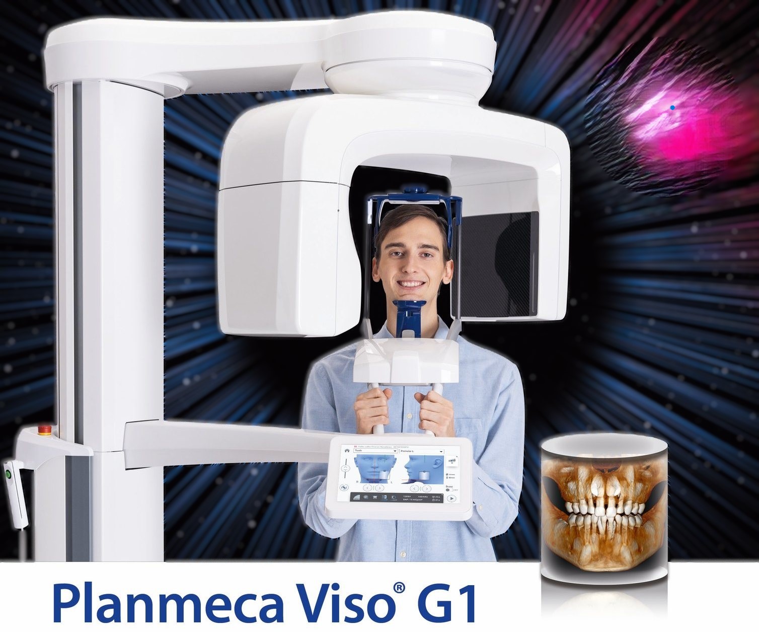
Next level CBCT imaging – go 3D with the Planmeca Viso® G1, a powerhouse of a unit covering volume sizes up to 11 x 11 cm. With all the essential imaging programs and top-of-the-line 3D imaging technology this is an X-ray device that easily meets your everyday dental imaging demands.
CBCT unit includes Planmeca Ultra Low Dose™ – a proprietary low dose 3D imaging protocol that enables CBCT imaging with an even lower patient radiation dose than standard panoramic imaging.
Planmeca Viso G1 can be equipped with a Planmeca ProCeph™ A one-shot cephalostat. The short acquisition time eliminates the risk of patient movement, which is particularly beneficial when imaging young children and other patients with a tendency to move during imaging.
Precision Meets Care: The Power of CBCT Imaging
In modern dentistry, we believe that you cannot treat what you cannot see. While traditional 2D X-rays have been the standard for decades and are still an important investigation they only tell part of the story.
To provide the highest level of clinical excellence, our practice utilises Cone Beam Computed Tomography (CBCT) in the appropriate cases. This advanced imaging technology allows us to view your oral anatomy in high-definition 3D, ensuring safer procedures and more predictable results. We were the first dental practice in the UK to have the Planmeca Visio G1 scanner that now employs AI technology.
What is a CBCT?
Unlike a standard dental X-ray, a CBCT scan uses a cone-shaped X-ray beam to capture hundreds of images in a single rotation. These images are reconstructed into a detailed 3D model of your teeth, jawbone, nerve pathways, and soft tissues.
Why 3D Imaging Matters for You
- Unparalleled Accuracy: We can see the exact height, width, and density of your bone, which is vital for complex procedures.
- Enhanced Safety: By identifying the precise location of nerves and sinuses, we minimise the risk of complications during surgery.
- Faster Appointments: A full scan takes less than 40 seconds, and the images are available for review instantly.
- Lower Radiation: CBCT technology is designed to provide maximum detail with significantly less radiation exposure than a conventional medical CT scan.
How We Use CBCT Technology
|
Procedure |
How CBCT Improves Your Outcome |
|
Dental Implants |
Allows for “Virtual Surgery” to place implants in the best position for long-term success. |
|
Root Canals |
Detects hidden canals or fractures that are invisible on 2D X-rays, preventing reinfection. |
|
Orthodontics |
Provides a clear view of impacted teeth and bone structure to map out the most efficient tooth movement. |
|
Wisdom teeth |
Allow us to assess the proximity of the lower wisdom tooth roots to the nerve that runs close to it in the lower jaw, and the complex root anatomy that may be associated with it |
|
TMJ Disorders |
Evaluates the jaw joint for signs of degeneration or misalignment. |
Frequently Asked Questions
Is the scan uncomfortable?
Not at all. You simply sit or stand still while the machine rotates around your head. There are no sensors to bite down on, making it a great option for patients with a strong gag reflex.
Is it worth the extra step?
Absolutely. Investing in a 3D scan at the beginning of your treatment when its relevant to do so, often saves time and money by preventing “surprises” during surgery and ensuring your treatment is done right the first time.
Experience the Difference of 3D Dentistry
Your health deserves the best technology available. During your next consultation, ask us how a CBCT scan may be of benefit to your dental care
CBCT Referral Form